
STURP - the Shroud of Turin Research
Project
In 1978 the Shroud was on public display for the first time since 1933. Over 3
million people passed through the Cathedral of St. John the Baptist to view it
behind bullet proof glass during the three weeks it was on display. Among the
pilgrims who view the Shroud is Karol, Cardinal Woytywa of Poland, shortly to
become Pope John Paul II.
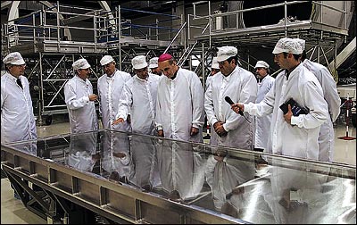
At the end of the exhibition, 40 scientists comprising the Shroud of Turin
Research Project (STURP), were allowed by the Cardinal of Turin to analyze the
Shroud for five continuous days (122 hours) working in shifts around the clock.
The STURP team is composed of scientists from universities, scientific
laboratories, and scientific industries. When the team’s results were published
in 1981, over 150,000 scientific hours had been employed in the research and
analysis.
The team's research continues today.
THE STURP
RESULTS
In 1981 the Shroud of Turin Research Project (STURP) team issued its Final
Report and the following official conclusions:
"No pigments, paints, dyes or stains have been found on the fibrils. X-ray,
fluorescence and microchemistry on the fibrils preclude the possibility of paint
being used as a method for creating the image. Ultra Violet and infrared
evaluation confirm these studies. Computer image enhancement and analysis by a
device known as a VP-8 image analyzer show that the image has unique,
three-dimensional information encoded in it.
Microchemical evaluation has indicated no evidence of any spices, oils, or any
biochemicals known to be produced by the body in life or in death. It is clear
that there has been a direct contact of the Shroud with a body, which explains
certain features such as scourge marks, as well as the blood. However, while
this type of contact might explain some of the features of the torso, it is
totally incapable of explaining the image of the face with the high resolution
that has been amply demonstrated by photography.
The basic problem from a scientific point of view is that some explanations
which might be tenable from a chemical point of view, are precluded by physics.
Contrariwise, certain physical explanations, which may be attractive are
completely precluded by the chemistry.
For an adequate explanation for the image of the Shroud, one must have an
explanation which is scientifically sound, from a physical, chemical, biological
and medical viewpoint. At the present, this type of solution does not appear to
be obtainable by the best efforts of the members of the Shroud Team.
Furthermore, experiments in physics and chemistry with old linen have failed to
reproduce adequately the phenomenon presented by the Shroud of Turin.
The scientific concensus is that the image was produced by something which
resulted in oxidation, dehydration and conjugation of the polysaccharide
structure of the microfibrils of the linen itself. Such changes can be
duplicated in the laboratory by certain chemical and physical processes. A
similar type of change in linen can be obtained by sulfuric acid or heat.
However, there are no chemical or physical methods known which can account for
the totality of the image, nor can any combination of physical, chemical,
biological or medical circumstances explain the image adequately.
Thus, the answer to the question of how the image was produced or what produced
the image remains, now, as it has in the past, a mystery.
We can conclude for now that the Shroud image is that of a real human form of a
scourged, crucified man. It is not the product of an artist. The blood stains
are composed of hemoglobin and also give a positive test for serum albumin.
The image is an ongoing mystery and until further chemical studies are made,
perhaps by this group of scientists, or perhaps by some scientists in the
future, the problem remains unsolved."
More information at: www.Shroud.com
Scientific
Investigation of the Shroud
Scientific investigation of
the Shroud of Turin began in 1898 when an amateur
photographer named Secondo Pia took the first photograph
of the Shroud and found to his amazement that his
negative was a high resolution positive image, which
meant that the image on the Shroud was a high resolution
negative image. This implied that it could not be
a painting since artists cannot accurately produce a
negative image because they never see one.
Subsequent investigation of the wounds observed on the
Shroud by experts in anatomy and medicine led them to
conclude that the images and blood marks on the Shroud
were in some way the result of a real human body that
had been wrapped in the Shroud. In 1976, using a
VP-8 image analyzer, it was discovered that there is 3D
or topographical information in the image on the Shroud
related to the body-to-cloth vertical distance.
Since such information does not exist in any painting or
photograph, this indicated that the image on the Shroud
could not be a painting or photograph. This
motivated scientists at leading national laboratories
and research facilities in the United States to form the
Shroud of Turin Research Project (STURP) to apply the
best scientific methods and equipment to determine how
the image on the Shroud was formed. About 24 of
their team went to Turin in 1978 where they were allowed
five days, 24 hours a day, to perform non-destructive
testing on the Shroud. The STURP investigation
found that:
·
The image has no pigment, no carrier, no brush strokes,
no clumping of material between the fibers or threads,
no cracking due to centuries of folding or rolling the
Shroud, and no stiffening of the cloth. This means
that the image could not be due to paint, dye, or stain.
·
There is no capillarity (soaking up of a liquid) of the
discoloration in the fibers or threads, so the image
could not be due to application of a liquid such as an
acid or a chemical in a liquid state.
·
The image is not luminescent under ultra-violet light.
This means that the image could not be due to a scorch
from contact of a hot object with the cloth.
·
The image is only visible in front lighting. It is
not visible in back lighting. From this, the STURP
team concluded that the image does not result from any
substance placed on the cloth, which means that the
image could not be a rubbing, a dusting, or a print.
·
A typical thread contains about 100 to 200 fibers.
The image is caused discoloration of only the top one or
two layers of fibers in a thread.
·
On a discolored fiber, the discoloration is located on
the outside circumference of the fiber, usually 360
degrees around the fiber. The thickness of this
discolored layer is about 0.2 microns, which is less
than a wavelength of light, and only a small fraction of
the 15 to 20-micron diameter of a fiber. The
inside of the fiber is not discolored.
·
The discoloration of any fibers in the image results
from a change in the electron bonding of the carbon
atoms that were already in the cellulose molecule.
This change in the electron bonding of the carbon atoms
is equivalent to a dehydration and oxidation of the
cellulose molecule. But how can this change in the
electron bonding of the carbon atoms be accomplished to
create an image of a crucified man?
More information at: http://www.shroudresearch.net